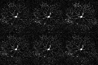
A starburst amacrine cell filled with dye through a patch pipette and visualized under a two photon microscope

1,729 retinal cells in the ganglion cell layer filled with Oregon Green calcium dye

A whole-retina antibody stain reveals the dorsal-ventral gradients of the two visual opsins in mice (S-opsin: magenta, M-opsin: green)

Immature retina cross-section - glial cells in green, glutamatergic synapses in pink - see Rosa, Bos et al, Elife, 2015

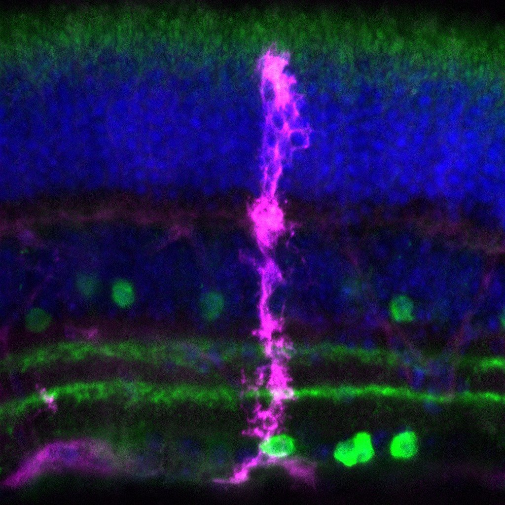
Adult retina cross-section - glial cells in green, glutamatergic synapses in pink -- see Rosa, Bos et al, Elife, 2015

Schematic of retinal projections influenced by retinal waves - see review by Kirkby et al, Neuron, 2013

Determining role of AII amacrine cells in retinal waves -- see Firl et al, J. Neurosci, 2014

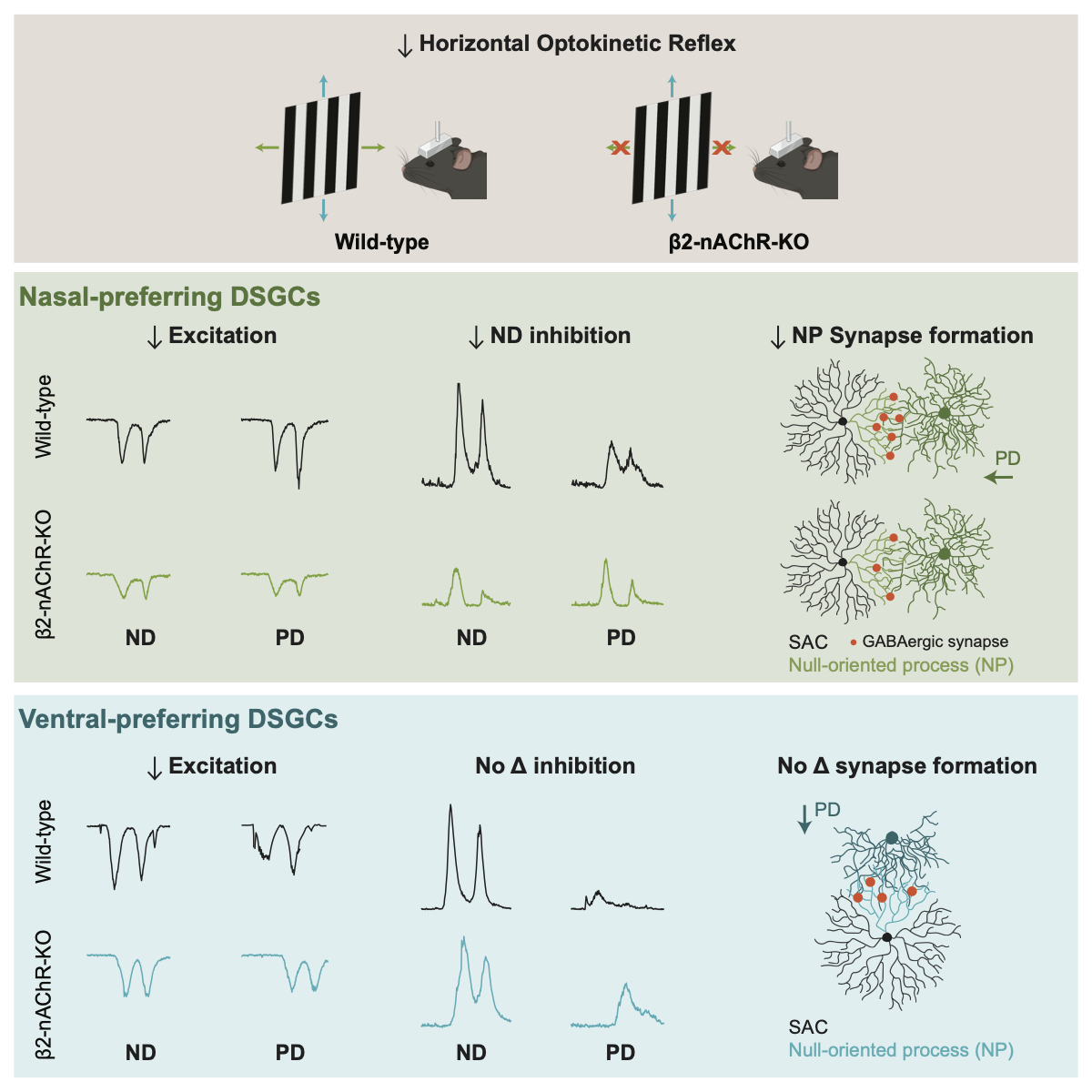
Paired whole cell voltage clamp recordings from a starburst amacrine cell and direction selective ganglion cell. See Morrie and Feller, J. Neurosci, 2015

Reconstructions of starburst-direction selective ganglion cell pair

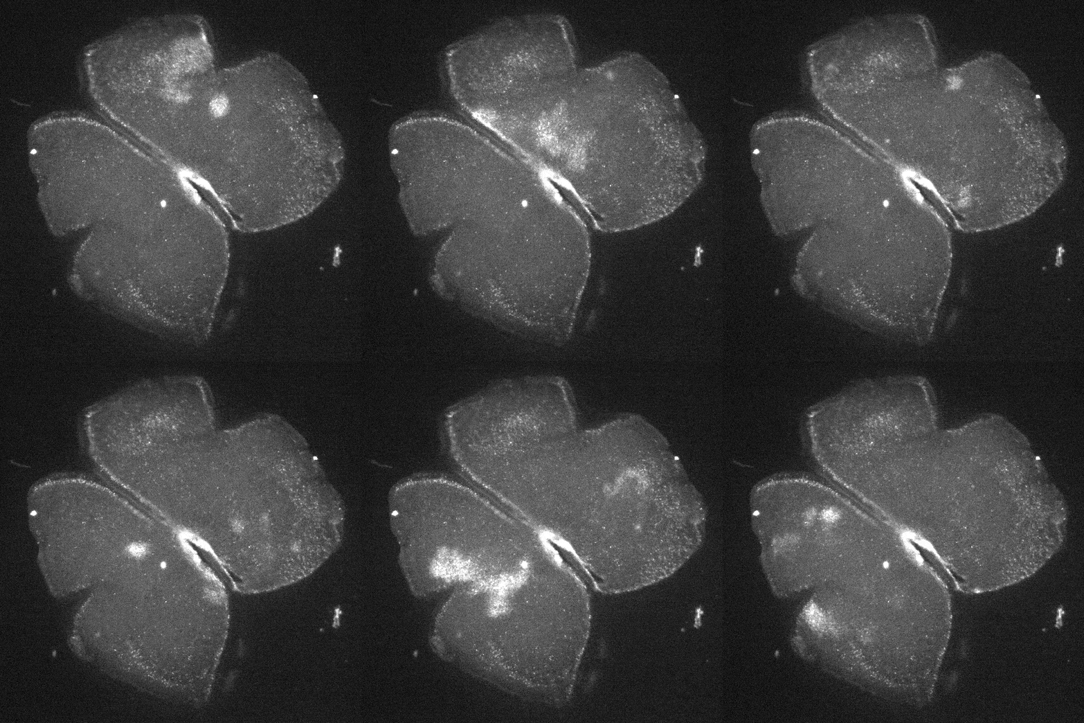
Imaging spillover of acetylcholine during retinal waves using CNiFERs -- see Ford et al, J. Neurosci, 2012

Synaptic currents evoked by light in Off-SAC -- see Vlasits et al, Neuron, 2014

Neurobiotin-filled ipRGC with gap junction coupled cells (magenta), and dopaminergic amacrine cells (green) in a P6 retina


Two-photon calcium imaging with resonance scanner
We study the functional development and organization of neural circuits in the retina.
We are part of the
Department of Molecular & Cell Biology,
Helen Wills Neuroscience Institute,
Biophysics Graduate Program
Vision Science Graduate Program at UC Berkeley