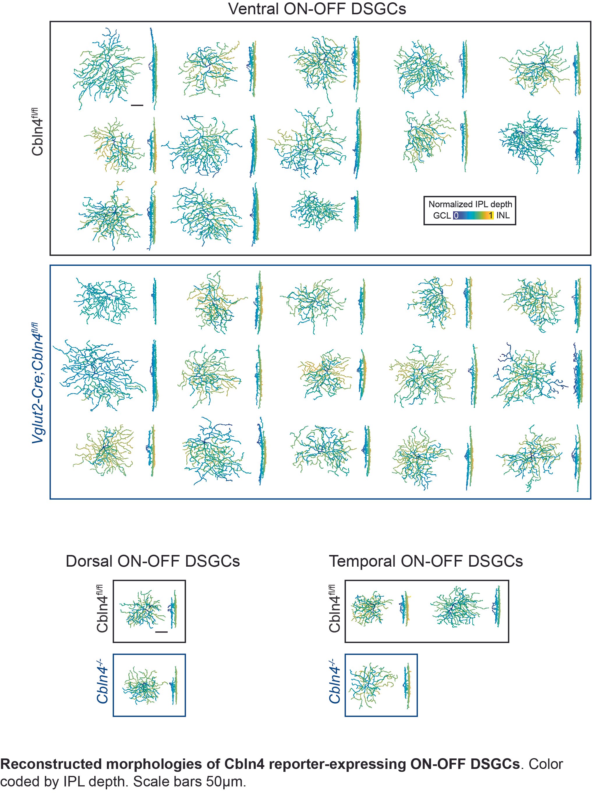
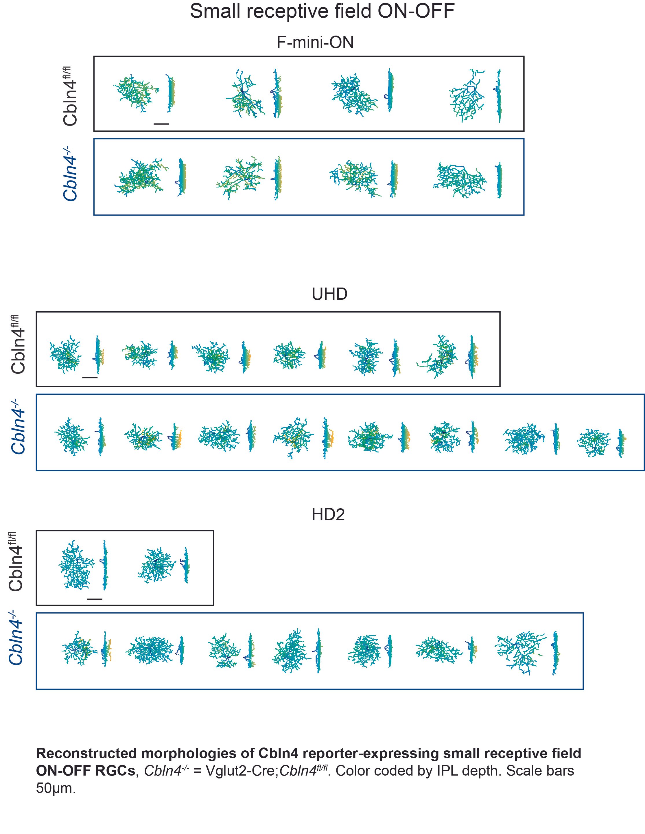
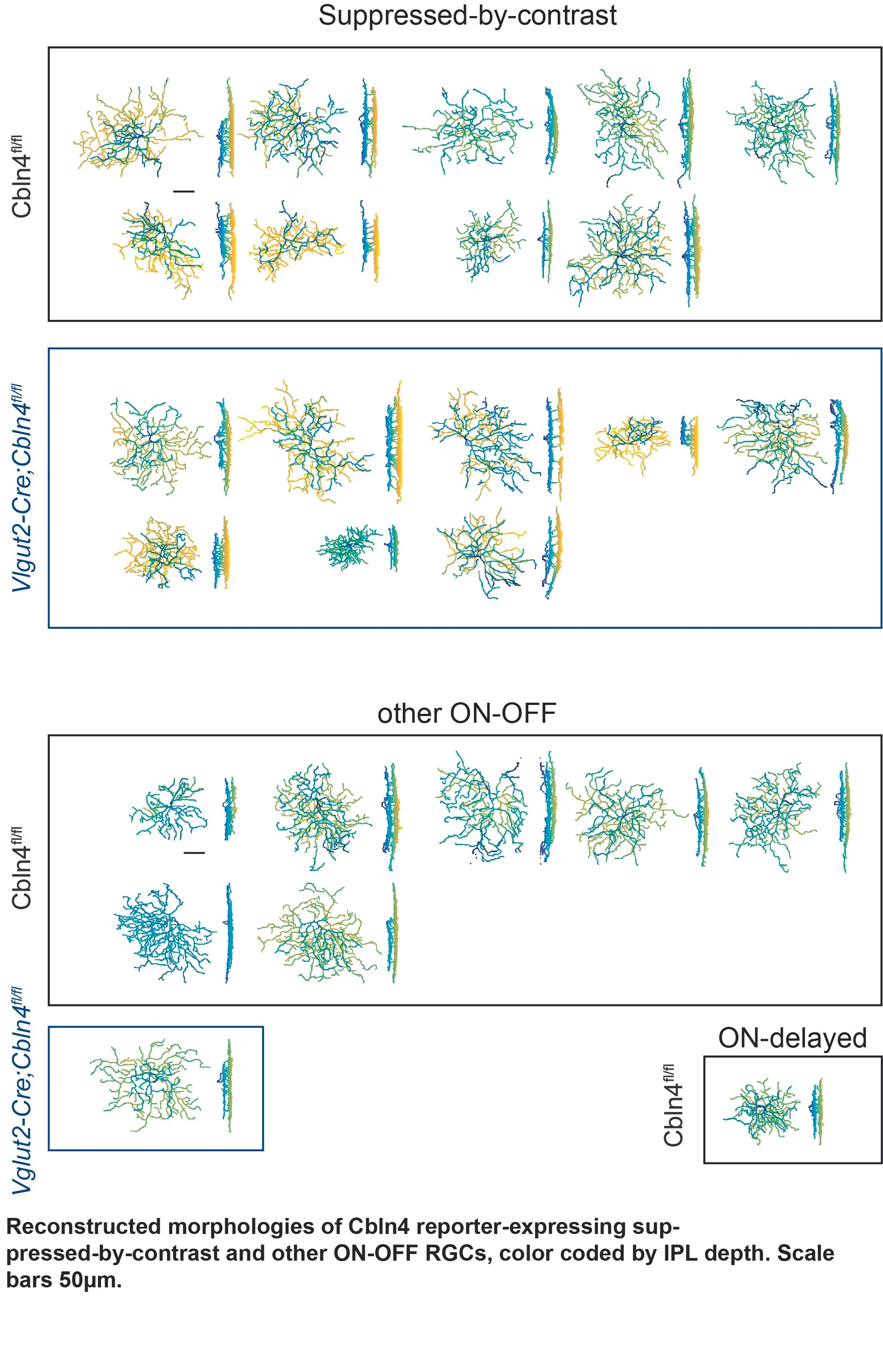
Reconstructions of Cbln4 expressing retinal ganglion cells - Supplement to Figure 8 of Tworig et al, 2023

Movie acquired with macroscope from P11 mouse expressing GCamP6s under the Vglut2 promoter. FOV is 5mm/side. Speed: 5x real time.
A TrpC 6/7 KO mouse exhibiting reduced photoaversion. Related to Figure 1, Cajal-Holme et al, 2022.
VIDEO here :
Compared to a wildtype pup (left column; 1:1 C129:C57BL6/J), a TrpC 6/7 KO pup (right column) exhibits reduced photoaversion. “Light on” text indicates the onset of the light stimulus (470 nm). Rows labels indicate the type of trial. Dark: no light stimulus. 7 and 9: 10^7 and 10^9 photons/ μm^2/sec at the eyelid, respectively, corresponding to ~10^5 or 10^7 photons / μm^2/sec at the retina. The flash at stimulus onset is from a 940 nm LED, to which melanopsin is ~10^12-fold less sensitive (Emanuel et al., 2015). Movies are synchronized for comparison, but each trial was conducted separately. Original acquisition at 28 Hz, playback at 5x real time.
M1 ipRGCs exempt from retinal waves. Related to Figure 6, Caval-Holme et al, J. Neuroscience, 2022
Video here: Two-photon Ca2+ imaging of retinal waves within the ganglion cell layer of a retina acutely dissected from a P8 wildtype (1:1 C129:C57BL6/J) mouse. Red outlines: somas of M1 ipRGCs that exhibited robust Ca2+responses during a light stimulus (first recording epoch) but not during retinal waves (latter three recording epochs). Note that other light-responsive neurons exhibit responses to waves. Original acquisition at 1.5 Hz. Playback at 5x. Scalebar is 50 µm.