Development of direction selectivity
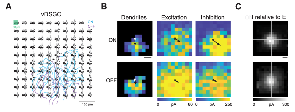
How are neural circuits wired up during development to perform computations? A classic neural computation is that of detecting the direction of motion of an object within the visual scene. Direction-selective cells are found in the retina and function as the first component of the reflex that stabilizes the visual image on the retina when an animal is in motion. Direction-selective ganglion cells respond strongly to an image moving in the preferred direction and weakly to an image moving in the opposite direction. This computation relies upon an asymmetric set of connections between inhibitory neurons onto direction selective cells as well as non-linearities in the cells themselves. We use a combination of approaches including imaging, opto- and pharmacogenetics and electrophysiology to determine the mechanisms that underlie the development of these asymmetric circuits. Ongoing projects, are based on recent findings that that blockade of retinal waves prevents the maturation of horizontal preferring DSGCs as well as a collaboration with Karthik Shekar’s lab to use scRNA-Seq methods to identify factors which intriuct the asymmetric wiring of inhibitory circuits.
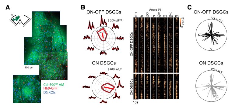
A. Tiriac, K. Bistrong, M. Pitcher, J. Tworig and M. B. Feller,. (2022) The influence of spontaneous and visual activity on the development of direction selectivity maps in mouse retina. Cell Reports, 38, 110225.
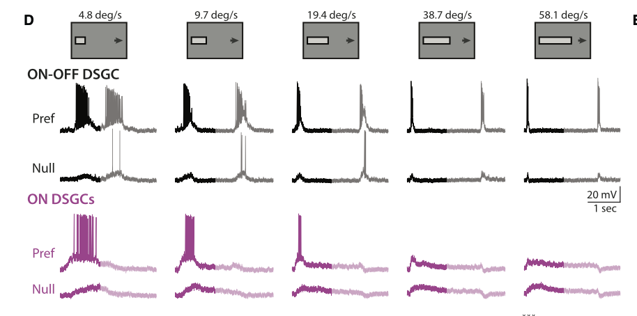
M. Summers and M. B. Feller (2022). Distinct Inhibitory Pathways Control Velocity and Directional Tuning in the Retina, Current Biology, 32(10):2130-2143 (highlighted in Dispatch by M. Manookin)
Other recent publications:
R. D. Morrie and M. B. Feller (2018). A dense starburst plexus is critical for generating direction selectivity, Current Biology, 28(8):1204-1212.
J. M. Rosa, R. D. Morrie, H. C. Baertsch and M. B. Feller (2016). The contributions of rod and cone pathways to retinal direction selectivity across light intensities and through development, Journal of Neuroscience, 36(37): 9683.
R. Bos, C. Gainer, M. B. Feller (2016). Role for visual experience in the development of the direction-selective circuits, Current Biology, 26(10):1367.
A. L. Vlasits, R. D. Morrie*, A. Bleckert*, A. Tran-Van-Minh*, C. F. Gainer, D. A. DiGregorio*, M. B. Feller* (2016). A role for synaptic input distribution in a dendritic computation of motion direction, Neuron 16;89(6):1317-30. (*equal contributions).
R. D. Morrie and M. B. Feller (2015), An asymmetric increase in inhibitory synapse number underlies the development of a direction selective circuit in the retina, Journal of Neuroscience 35(25):9281-6.
A. M. Hamby, J. M. Rosa, C-H Hsu and M. B. Feller (2015), CaV3.2 KO mice have altered retinal waves but normal direction selectivity, Visual Neuroscience, 32:E003.
M. El-Quessny and M. B. Feller. (2021) Dendrite morphology has limited impact on the spatial distribution of synaptic inputs onto retinal direction selective ganglion cells, eNeuro. 8(5): ENEURO.0261-21.2021.
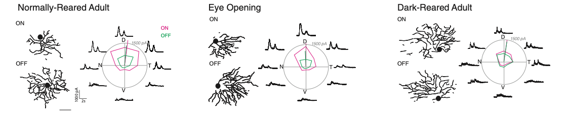
M. El-Quessny, K. Maanum and M. B. Feller. (2020) Visual experience instructs dendrite orientation but is not required for asymmetric wiring of the retinal direction selective circuit, Cell Reports, 31(13):107844
Reviews:
A. Tiriac and Feller, M. B. (2022) Roles of visually evoked and spontaneous activity in the development of retinal direction selectivity maps, Trends in Neuroscience, accepted for publication
M. Summers, M. El-Quessny and M. B. Feller, Retinal Mechanisms of Direction Selectivity (2021), Oxford Research Encyclopedia of Neuroscience, doi:10.1093/acrefore/9780190264086.013.356.
A. S. Mauss*, A.L. Vlasits** A. Borst#, M. B. Feller# (2017), “Visual Circuits for Direction Selectivity”, Annual Review Neuroscience, 40:211-230. (*,equal contributions)
R. D. Morrie and M. B. Feller (2016), “Development of synaptic connectivity in the retinal direction selective circuit”, Current Opinion in Neurobiology 40, 45-52.